| Revision as of 12:32, 27 November 2012 edit71.181.88.23 (talk)No edit summaryTag: repeating characters← Previous edit | Latest revision as of 20:21, 11 October 2024 edit undoA person of sorts (talk | contribs)Extended confirmed users857 edits Undid revision 1250672854 by 50.5.26.166 (talk)Tag: Undo | ||
| (887 intermediate revisions by more than 100 users not shown) | |||
| Line 1: | Line 1: | ||
| {{Short description|Skin and other protective organs}} | |||
| {{hatnote|"Integumentary " redirects here; in ], an integument refers to an outer membrane of an ], which later develops into the ], the ]}} | |||
| {{Redirect|Integumentary|the part of the female reproductive system of seed plants|Ovule}} | |||
| {{Infobox Anatomy | | |||
| {{Infobox anatomy | |||
| Name = Integumentary system |also known as the "skin system" | |||
| | Name = Integumentary system | |||
| | Image = Human_skin_structure.svg | |||
| '''GraySubject''' = | | |||
| | Caption = Cross-section of all skin layers | |||
| Image = | | |||
| | Width = | |||
| | Precursor = | |||
| | System = | |||
| Precursor = | | |||
| '''System''' = | | |||
| ''Artery'' = | | |||
| Vein = | | |||
| Nerve = | | |||
| Lymph = | | |||
| MeshName = | | |||
| MeshNumber = | | |||
| Code = ] H3.12.00.0.00001 | | |||
| }} | }} | ||
| The '''integumentary system''' is the ] that protects the body from damage, comprising the ] and its appendages<ref>{{MeshName|Integumentary+System}}</ref><ref>{{cite book | |||
| | last=Marieb | first=Elaine | coauthors=Katja Hoehn | |||
| | title=Human Anatomy & Physiology | |||
| | publisher=Pearson Benjamin Cummings | |||
| | year=2007 | |||
| | edition=7th | |||
| | page=142 | |||
| }}</ref> (including ], ], ], ]s, and ]). The integumentary system has a variety of functions; it may serve to waterproof, cushion, and protect the deeper tissues, excrete wastes, and regulate ], and is the attachment site for ]s to detect pain, sensation, pressure, and temperature. In most terrestrial vertebrates with significant exposure to sunlight, the integumentary system also provides for ] synthesis. | |||
| The '''integumentary system''' is the set of organs forming the outermost layer of an animal's body. It comprises the ] and its appendages, which act as a physical barrier between the external environment and the internal environment that it serves to protect and maintain the body of the animal. Mainly it is the body's outer skin. | |||
| ==Layers of the skin== | |||
| The integumentary system is the largest of the body's ]. In humans, this system accounts for about 12 to 15 percent of total body weight and covers 1.5-2m<sup>2</sup> of surface area.<ref>Martini & Nath: "Fundamentals of Anatomy & Physiology" 8th Edition, pp.158,Pearson Education, 2009</ref> It distinguishes, separates, and protects the organism from its surroundings. Small-bodied invertebrates of aquatic or contifdfd edcgdshgdsh dgyfsugfygsy sygysdyfsnually moist habitats ] using the outer layer (integument). This gas exchange system, where gases simply diffuse into and out of the ], is called '''integumentary exchange'''. | |||
| The integumentary system includes ], ], ], ], ], ]s, and ]. It has a variety of additional functions: it may serve to maintain water balance, protect the deeper tissues, excrete wastes, and regulate ], and is the attachment site for ]s which detect pain, sensation, pressure, and temperature. | |||
| The human skin (integument) is composed of a minimum of 3 major layers of tissue: the ]; ]; and ]. The epidermis forms the outermost layer, providing the initial barrier to the external environment. Beneath this, the dermis comprises two sections, the papillary and reticular layers, and contains connective tissues, vessels, glands, follicles, hair roots, sensory nerve endings, and muscular tissue.<ref name="ageing skin"></ref> The deepest layer is the hypodermis, which is primarily made up of ]. Substantial collagen bundles anchor the dermis to the hypodermis in a way that permits most areas of the skin to move freely over the deeper tissue layers.<ref>{{cite web|last=Pratt|first=Rebecca|title=Integument|url=http://www.anatomyone.com/a/integument/|work=AnatomyOne|publisher=Amirsys, Inc|accessdate=2012-09-28}}</ref> | |||
| == |
==Structure== | ||
| === Skin === | |||
| {{Main|Epidermis (skin)}} | |||
| {{Main|Skin}} | |||
| This is the top layer of skin made up of ]. It does not contain blood vessels. Its main function is protection, absorption of nutrients, and homeostasis. In structure, it consists of a keratinized stratified ] comprising four types of cells: ], ], ], and ]. The major cell of the epidermis is the keratinocyte, which produces keratin. ] is a fibrous protein that aids in protection. Keratin is also a water-proofing protein. Millions of dead keratinocytes rub off daily. The majority of the skin on the body is keratinized, meaning ]. The only skin on the body that is non-keratinized is the lining of skin on the inside of the mouth. Non-keratinized cells allow water to "stay" atop the structure. | |||
| The skin is one of the largest organs of the body. In humans, it accounts for about 12 to 15 percent of total body weight and covers 1.5 to 2 m<sup>2</sup> of surface area.<ref>{{cite book |last1=Martini |first1=Frederic |last2=Nath |first2=Judi L. |title=Fundamentals of anatomy & physiology |date=2009 |publisher=Pearson/Benjamin Cummings |location=San Francisco |isbn=978-0321505897 |page=158 |edition=8th}}</ref> | |||
| ] | |||
| The skin (integument) is a composite organ, made up of at least two major layers of tissue: the ] and the ].<ref name="Kardong2019">{{cite book |last1=Kardong |first1=Kenneth V. |title=Vertebrates : comparative anatomy, function, evolution |date=2019 |location=New York, NY |isbn=978-1-259-70091-0 |pages=212–214 |edition=Eighth}}</ref> The epidermis is the outermost layer, providing the initial barrier to the external environment. It is separated from the dermis by the ] (] and ]). The epidermis contains ]s and gives color to the skin. The deepest layer of the epidermis also contains ]s. Beneath this, the dermis comprises two sections, the papillary and reticular layers, and contains ]s, vessels, glands, follicles, ]s, sensory nerve endings, and muscular tissue.<ref name="aging skin">{{cite web |title=The Ageing Skin – Part 1 – Structure of Skin |url=http://pharmaxchange.info/press/2011/03/the-ageing-skin-part-1-structure-of-skin-and-introduction |website=pharmaxchange.info|date=4 March 2011 }}</ref> | |||
| The protein keratin stiffens epidermal tissue to form ]s. Nails grow from thin area called the ]; growth of nails is 1 mm per week on average. The ] is the crescent-shape area at the base of the nail, this is a lighter color as it mixes with the matrix cells. | |||
| Between the integument and the deep body musculature there is a transitional subcutaneous zone made up of very loose connective and ], the ]. Substantial ] bundles anchor the dermis to the hypodermis in a way that permits most areas of the skin to move freely over the deeper tissue layers.<ref>{{cite web|last=Pratt|first=Rebecca|title=Integument|url=http://www.anatomyone.com/a/integument/|work=AnatomyOne|publisher=Amirsys, Inc|access-date=2012-09-28|archive-date=2013-10-20|archive-url=https://web.archive.org/web/20131020130128/http://www.anatomyone.com/a/integument/|url-status=dead}}</ref> | |||
| ===Dermis=== | |||
| ====Epidermis==== | |||
| {{Main|Epidermis}} | |||
| ] | |||
| The ] is the strong, superficial layer that serves as the first line of protection against the outer environment. The human epidermis is composed of ], which further break down into four to five layers: the ], ], ] and ]. Where the skin is thicker, such as in the palms and soles, there is an extra layer of skin between the stratum corneum and the stratum granulosum, called the ]. The epidermis is regenerated from the stem cells found in the basal layer that develop into the corneum. The epidermis itself is devoid of blood supply and draws its nutrition from its underlying dermis.<ref name="statpearls2"/> | |||
| Its main functions are protection, absorption of nutrients, and ]. In structure, it consists of a keratinized stratified ]; four types of cells: ], ], ], and ]. | |||
| The predominant cell ], which produces ], a fibrous protein that aids in skin protection, is responsible for the formation of the epidermal water barrier by making and secreting ]s.<ref name="statpearls">{{cite book |last1=Yousef |first1=Hani |last2=Alhajj |first2=Mandy |last3=Sharma |first3=Sandeep |title=StatPearls |publisher=StatPearls Publishing |url=https://www.ncbi.nlm.nih.gov/books/NBK470464/ |chapter=Anatomy, Skin (Integument), Epidermis|year=2022 |pmid=29262154 }}</ref> <!--An overwhelming amount of keratin can cause disease by giving rise to eruptions from the skin that will protrude outwards and lead to infection.{{Citation needed|date=March 2017}}--> The majority of the skin on the human body is keratinized, with the exception of the lining of ]s, such as the inside of the mouth. Non-keratinized cells allow water to "stay" atop the structure. | |||
| The protein keratin stiffens epidermal tissue to form ]s. Nails grow from a thin area called the ] at an average of 1 mm per week. The ] is the crescent-shape area at the base of the nail, lighter in color as it mixes with matrix cells. Only ]s have nails. In other vertebrates, the keratinizing system at the terminus of each digit produces claws or hooves.<ref name="Kardong2019"/> | |||
| The epidermis of vertebrates is surrounded by two kinds of coverings, which are produced by the epidermis itself. In ] and aquatic ]s, it is a thin mucus layer that is constantly being replaced. In terrestrial vertebrates, it is the ] (dead keratinized cells). The epidermis is, to some degree, glandular in all vertebrates, but more so in ] and ]s. Multicellular epidermal glands penetrate the dermis, where they are surrounded by blood capillaries that provide nutrients and, in the case of endocrine glands, transport their products.<ref>{{cite journal |last1=Quay |first1=Wilbur B. |title=Integument and the Environment Glandular Composition, Function, and Evolution |journal=Integrative and Comparative Biology |date=1 February 1972 |volume=12 |issue=1 |pages=95–108 |url=https://academic.oup.com/icb/article/12/1/95/2107657}}</ref> | |||
| {{Clear|left}} | |||
| ====Dermis==== | |||
| {{Main|Dermis}} | {{Main|Dermis}} | ||
| The dermis is the |
The dermis is the underlying connective tissue layer that supports the epidermis. It is composed of dense irregular connective tissue and ] such as a collagen with ] arranged in a diffusely bundled and woven pattern. | ||
| The dermis has two layers: the papillary dermis and the reticular layer. The papillary layer is the superficial layer that forms finger-like projections into the epidermis (dermal papillae),<ref name="statpearls2">{{cite book |last1=Kim |first1=Joyce Y. |last2=Dao |first2=Harry |title=StatPearls |publisher=StatPearls Publishing |url=https://www.ncbi.nlm.nih.gov/books/NBK554386/ |chapter=Physiology, Integument|year=2022 |pmid=32119273 }}</ref> and consists of highly vascularized, loose connective tissue. The reticular layer is the deep layer of the dermis and consists of the dense irregular connective tissue. These layers serve to give elasticity to the integument, allowing stretching and conferring flexibility, while also resisting distortions, wrinkling, and sagging.<ref name="aging skin"/> The dermal layer provides a site for the endings of blood vessels and nerves. Many ] are also stored in this layer, as are the bases of integumental structures such as ], ], and ]. | |||
| ===Hypodermis=== | ===Hypodermis=== | ||
| {{Main| |
{{Main|Hypodermis}} | ||
| The hypodermis, otherwise known as the subcutaneous layer, is a layer beneath the skin. It invaginates into the dermis and is attached to the latter, immediately above it, by collagen and elastin fibers. It is essentially composed of a type of cell known as adipocytes, which are specialized in accumulating and storing fats. These cells are grouped together in lobules separated by connective tissue. | |||
| Also called the hypoderm, subcutaneous tissue, or superficial fascia and the bottom layer of the integumentary system in vertebrates (hypoderm and subcutaneous are from Greek and Latin words, respectively, for "beneath the skin"). Types of cells that are found in the hypodermis are fibroblasts, adipose cells, and macrophages. It is derived from the mesoderm, but unlike the dermis, it is not derived from the dermatome region of the mesoderm. In arthropods, the hypodermis is an epidermal layer of cells that secretes the chitinous cuticle. | |||
| The hypodermis acts as an energy reserve. The fats contained in the adipocytes can be put back into circulation, via the venous route, during intense effort or when there is a lack of energy-providing substances, and are then transformed into energy. The hypodermis participates, passively at least, in thermoregulation since fat is a heat insulator. | |||
| ==Functions== | ==Functions== | ||
| The integumentary system has multiple roles in ]. All body systems work in an interconnected manner to maintain the internal conditions essential to the function of the body. The skin has an important job of protecting the body and acts as the |
The integumentary system has multiple roles in ]. All body systems work in an interconnected manner to maintain the internal conditions essential to the function of the body. The skin has an important job of protecting the body and acts as the body's first line of defense against infection, temperature change, and other challenges to homeostasis.<ref>{{MeSH name|Integumentary+System}}</ref><ref>{{cite book | last=Marieb | first=Elaine |author2=Hoehn, Katja | title=Human Anatomy & Physiology | url=https://archive.org/details/humananatomyphys00mari_4 | url-access=registration | publisher=] | year=2007 | edition=7th | page=| isbn=9780805359107 }}</ref> | ||
| Its main functions include: | |||
| *Protect the |
*Protect the body's internal living ]s and organs | ||
| *Protect against invasion by ] organisms | *Protect against invasion by ] organisms | ||
| *Protect the body from ] | *Protect the body from ] | ||
| Line 57: | Line 63: | ||
| *Protect the body against ]s by secreting melanin | *Protect the body against ]s by secreting melanin | ||
| *Generate ] through exposure to ] ] | *Generate ] through exposure to ] ] | ||
| *Store ], ], glucose, |
*Store ], ], glucose, vitamin D | ||
| *Maintenance of the body form | *Maintenance of the body form | ||
| *Formation of new cells from stratum germinativum to repair minor injuries | *Formation of new cells from stratum germinativum to repair minor injuries | ||
| *Protect from ] rays. | |||
| *Aid in physical examination as color of the skin may indicate many conditions e.g. it becomes yellowish in jaundice | |||
| *Regulates body temperature | |||
| *It distinguishes, separates, and protects the organism from its surroundings. | |||
| Small-bodied invertebrates of aquatic or continually moist habitats ] using the outer layer (integument). This gas exchange system, where gases simply diffuse into and out of the ], is called '''integumentary exchange'''. | |||
| ==Clinical significance== | |||
| {{For|a comprehensive list|List of cutaneous conditions}} | |||
| ==Diseases and injuries== | |||
| Possible diseases and injuries to the human integumentary system include: | Possible diseases and injuries to the human integumentary system include: | ||
| {{Div col|colwidth=10em}} | |||
| *] | |||
| *] | * ] | ||
| *] | * ] | ||
| *] | * ] | ||
| *] | * ] | ||
| *] | * ] | ||
| *] | * ] | ||
| *] | * ] | ||
| *] | * ] | ||
| *] | * ] | ||
| * ], commonly called cold sores | |||
| *] | |||
| *] | * ] | ||
| *] | * ] | ||
| * ] | |||
| *] | |||
| *] | * ] | ||
| * ] | |||
| * ] | |||
| * ] | |||
| * ] | |||
| {{Div col end}} | |||
| ==References== | ==References== | ||
| {{Reflist}} | {{Reflist}} | ||
| ==External links== | |||
| {{Organ systems}} | |||
| {{Wikibooks|Human Physiology|Integumentary System}} | |||
| {{Library resources box | |||
| |by=no | |||
| |onlinebooks=no | |||
| |others=no | |||
| |about=yes | |||
| |label=Integumentary System}} | |||
| {{Human systems and organs}} | |||
| {{Integumentary system}} | {{Integumentary system}} | ||
| {{Authority control}} | |||
| {{DEFAULTSORT:Integumentary System}} | {{DEFAULTSORT:Integumentary System}} | ||
| ] | ] | ||
| ] | ] | ||
| ] | |||
| ] | |||
| ] | |||
| ] | |||
| ] | |||
| ] | |||
| ] | |||
| ] | |||
| ] | |||
| ] | |||
| ] | |||
| ] | |||
| ] | |||
| ] | |||
| ] | |||
| ] | |||
| ] | |||
| ] | |||
| ] | |||
| ] | |||
| ] | |||
| ] | |||
| ] | |||
| ] | |||
| ] | |||
| ] | |||
| ] | |||
| ] | |||
| ] | |||
| ] | |||
| ] | |||
| ] | |||
| ] | |||
| ] | |||
| ] | |||
| ] | |||
| ] | |||
| ] | |||
| ] | |||
| ] | |||
| ] | |||
| ] | |||
| ] | |||
| ] | |||
| ] | |||
| ] | |||
| ] | |||
| ] | |||
| ] | |||
| ] | |||
| ] | |||
| ] | |||
| ] | |||
| ] | |||
Latest revision as of 20:21, 11 October 2024
Skin and other protective organs "Integumentary" redirects here. For the part of the female reproductive system of seed plants, see Ovule.| Integumentary system | |
|---|---|
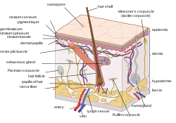
 Cross-section of all skin layers Cross-section of all skin layers | |
| Identifiers | |
| MeSH | D034582 |
| TA98 | A16.0.00.001 |
| TA2 | 7040 |
| TH | H3.12.00.0.00001 |
| FMA | 72979 |
| Anatomical terminology[edit on Wikidata] | |
The integumentary system is the set of organs forming the outermost layer of an animal's body. It comprises the skin and its appendages, which act as a physical barrier between the external environment and the internal environment that it serves to protect and maintain the body of the animal. Mainly it is the body's outer skin.
The integumentary system includes skin, hair, scales, feathers, hooves, claws, and nails. It has a variety of additional functions: it may serve to maintain water balance, protect the deeper tissues, excrete wastes, and regulate body temperature, and is the attachment site for sensory receptors which detect pain, sensation, pressure, and temperature.
Structure
Skin
Main article: SkinThe skin is one of the largest organs of the body. In humans, it accounts for about 12 to 15 percent of total body weight and covers 1.5 to 2 m of surface area.

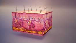
The skin (integument) is a composite organ, made up of at least two major layers of tissue: the epidermis and the dermis. The epidermis is the outermost layer, providing the initial barrier to the external environment. It is separated from the dermis by the basement membrane (basal lamina and reticular lamina). The epidermis contains melanocytes and gives color to the skin. The deepest layer of the epidermis also contains nerve endings. Beneath this, the dermis comprises two sections, the papillary and reticular layers, and contains connective tissues, vessels, glands, follicles, hair roots, sensory nerve endings, and muscular tissue.
Between the integument and the deep body musculature there is a transitional subcutaneous zone made up of very loose connective and adipose tissue, the hypodermis. Substantial collagen bundles anchor the dermis to the hypodermis in a way that permits most areas of the skin to move freely over the deeper tissue layers.
Epidermis
Main article: Epidermis
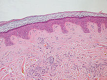
The epidermis is the strong, superficial layer that serves as the first line of protection against the outer environment. The human epidermis is composed of stratified squamous epithelial cells, which further break down into four to five layers: the stratum corneum, stratum granulosum, stratum spinosum and stratum basale. Where the skin is thicker, such as in the palms and soles, there is an extra layer of skin between the stratum corneum and the stratum granulosum, called the stratum lucidum. The epidermis is regenerated from the stem cells found in the basal layer that develop into the corneum. The epidermis itself is devoid of blood supply and draws its nutrition from its underlying dermis.
Its main functions are protection, absorption of nutrients, and homeostasis. In structure, it consists of a keratinized stratified squamous epithelium; four types of cells: keratinocytes, melanocytes, Merkel cells, and Langerhans cells.
The predominant cell keratinocyte, which produces keratin, a fibrous protein that aids in skin protection, is responsible for the formation of the epidermal water barrier by making and secreting lipids. The majority of the skin on the human body is keratinized, with the exception of the lining of mucous membranes, such as the inside of the mouth. Non-keratinized cells allow water to "stay" atop the structure.
The protein keratin stiffens epidermal tissue to form fingernails. Nails grow from a thin area called the nail matrix at an average of 1 mm per week. The lunula is the crescent-shape area at the base of the nail, lighter in color as it mixes with matrix cells. Only primates have nails. In other vertebrates, the keratinizing system at the terminus of each digit produces claws or hooves.
The epidermis of vertebrates is surrounded by two kinds of coverings, which are produced by the epidermis itself. In fish and aquatic amphibians, it is a thin mucus layer that is constantly being replaced. In terrestrial vertebrates, it is the stratum corneum (dead keratinized cells). The epidermis is, to some degree, glandular in all vertebrates, but more so in fish and amphibians. Multicellular epidermal glands penetrate the dermis, where they are surrounded by blood capillaries that provide nutrients and, in the case of endocrine glands, transport their products.
Dermis
Main article: DermisThe dermis is the underlying connective tissue layer that supports the epidermis. It is composed of dense irregular connective tissue and areolar connective tissue such as a collagen with elastin arranged in a diffusely bundled and woven pattern.
The dermis has two layers: the papillary dermis and the reticular layer. The papillary layer is the superficial layer that forms finger-like projections into the epidermis (dermal papillae), and consists of highly vascularized, loose connective tissue. The reticular layer is the deep layer of the dermis and consists of the dense irregular connective tissue. These layers serve to give elasticity to the integument, allowing stretching and conferring flexibility, while also resisting distortions, wrinkling, and sagging. The dermal layer provides a site for the endings of blood vessels and nerves. Many chromatophores are also stored in this layer, as are the bases of integumental structures such as hair, feathers, and glands.
Hypodermis
Main article: HypodermisThe hypodermis, otherwise known as the subcutaneous layer, is a layer beneath the skin. It invaginates into the dermis and is attached to the latter, immediately above it, by collagen and elastin fibers. It is essentially composed of a type of cell known as adipocytes, which are specialized in accumulating and storing fats. These cells are grouped together in lobules separated by connective tissue.
The hypodermis acts as an energy reserve. The fats contained in the adipocytes can be put back into circulation, via the venous route, during intense effort or when there is a lack of energy-providing substances, and are then transformed into energy. The hypodermis participates, passively at least, in thermoregulation since fat is a heat insulator.
Functions
The integumentary system has multiple roles in maintaining the body's equilibrium. All body systems work in an interconnected manner to maintain the internal conditions essential to the function of the body. The skin has an important job of protecting the body and acts as the body's first line of defense against infection, temperature change, and other challenges to homeostasis.
Its main functions include:
- Protect the body's internal living tissues and organs
- Protect against invasion by infectious organisms
- Protect the body from dehydration
- Protect the body against abrupt changes in temperature, maintain homeostasis
- Help excrete waste materials through perspiration
- Act as a receptor for touch, pressure, pain, heat, and cold (see Somatosensory system)
- Protect the body against sunburns by secreting melanin
- Generate vitamin D through exposure to ultraviolet light
- Store water, fat, glucose, vitamin D
- Maintenance of the body form
- Formation of new cells from stratum germinativum to repair minor injuries
- Protect from UV rays.
- Regulates body temperature
- It distinguishes, separates, and protects the organism from its surroundings.
Small-bodied invertebrates of aquatic or continually moist habitats respire using the outer layer (integument). This gas exchange system, where gases simply diffuse into and out of the interstitial fluid, is called integumentary exchange.
Clinical significance
For a comprehensive list, see List of cutaneous conditions.Possible diseases and injuries to the human integumentary system include:
- Rash
- Yeast
- Athlete's foot
- Infection
- Sunburn
- Skin cancer
- Albinism
- Acne
- Herpes
- Herpes labialis, commonly called cold sores
- Impetigo
- Rubella
- Cancer
- Psoriasis
- Rabies
- Rosacea
- Atopic dermatitis
- Eczema
References
- Martini, Frederic; Nath, Judi L. (2009). Fundamentals of anatomy & physiology (8th ed.). San Francisco: Pearson/Benjamin Cummings. p. 158. ISBN 978-0321505897.
- ^ Kardong, Kenneth V. (2019). Vertebrates : comparative anatomy, function, evolution (Eighth ed.). New York, NY. pp. 212–214. ISBN 978-1-259-70091-0.
{{cite book}}: CS1 maint: location missing publisher (link) - ^ "The Ageing Skin – Part 1 – Structure of Skin". pharmaxchange.info. 4 March 2011.
- Pratt, Rebecca. "Integument". AnatomyOne. Amirsys, Inc. Archived from the original on 2013-10-20. Retrieved 2012-09-28.
- ^ Kim, Joyce Y.; Dao, Harry (2022). "Physiology, Integument". StatPearls. StatPearls Publishing. PMID 32119273.
- Yousef, Hani; Alhajj, Mandy; Sharma, Sandeep (2022). "Anatomy, Skin (Integument), Epidermis". StatPearls. StatPearls Publishing. PMID 29262154.
- Quay, Wilbur B. (1 February 1972). "Integument and the Environment Glandular Composition, Function, and Evolution". Integrative and Comparative Biology. 12 (1): 95–108.
- Integumentary+System at the U.S. National Library of Medicine Medical Subject Headings (MeSH)
- Marieb, Elaine; Hoehn, Katja (2007). Human Anatomy & Physiology (7th ed.). Pearson Benjamin Cummings. p. 142. ISBN 9780805359107.
External links
Library resources aboutIntegumentary System
| Skin and related structures | |||||||||||||||
|---|---|---|---|---|---|---|---|---|---|---|---|---|---|---|---|
| Skin |
| ||||||||||||||
| Subcutaneous tissue | |||||||||||||||
| Adnexa |
| ||||||||||||||