Medical condition
| Bladder cancer | |
|---|---|
 | |
| Transitional cell carcinoma of the bladder. The white in the bladder is contrast. | |
| Specialty | Oncology, urology |
| Symptoms | Blood in the urine |
| Usual onset | Age 65 or older |
| Types | Non-muscle-invasive bladder cancer (NMIBC), muscle-invasive bladder cancer (MIBC), metastatic bladder cancer |
| Risk factors | Tobacco smoking, exposure to certain toxic chemicals, schistosomiasis infection |
| Diagnostic method | Cystoscopy with tissue biopsy |
| Treatment | Transurethral resection (TURBT), chemotherapy, BCG vaccine, immunotherapy, radical cystectomy, molecular-targeted therapies |
| Prognosis | Five-year survival rates range from 5% to 96% depending on the stage |
| Frequency | Around 500,000 each year |
| Deaths | Around 200,000 each year |
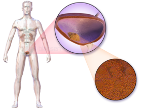
Bladder cancer is the abnormal growth of cells on the bladder. These cells, which originate in the urothelium, can grow to form a tumor, which eventually spreads, damaging the bladder and other organs. Most people with bladder cancer are diagnosed after noticing blood in their urine. Those suspected of having bladder cancer typically have their bladder inspected by cystoscopy, a procedure where a thin medical camera is inserted through the urethra. Suspected tumors are removed and examined by a pathologist to determine if they are cancerous. Based on how far the tumor has spread, the cancer case is assigned a stage 0 to 4; a higher stage indicates a more widespread and dangerous disease.
Those whose bladder tumors are completely contained within the bladder have the best prognoses. These tumors are typically surgically removed, and the person is treated with chemotherapy or an immune-stimulating therapy – either the BCG vaccine or one of several alternative immune stimulants. Those whose tumors continue to grow, or whose tumors have penetrated the bladder muscle, are often treated with radical cystectomy, the surgical removal of the bladder and nearby genital organs. Those whose tumors have spread beyond the bladder have the worst prognoses and on average a person survives a year from diagnosis. These people are treated with chemotherapy and immune checkpoint inhibitors, followed by the antibody drug conjugate therapy enfortumab vedotin.
Around 500,000 people are diagnosed with bladder cancer each year, and 200,000 die of the disease. The risk of bladder cancer increases with age and the average age at diagnosis is 73. Tobacco smoking is the greatest contributor to bladder cancer risk, and causes around half of bladder cancer cases. Exposure to certain toxic chemicals or the flatworm Schistosoma haematobium, which infects the bladder, also increases the risk.
Signs and symptoms

The most common symptom of bladder cancer is visible blood in the urine (haematuria) despite painless urination. This affects around 75% of people eventually diagnosed with the disease. Some instead have "microscopic haematuria" – small amounts of blood in the urine that can only be seen under a microscope during urinalysis – pain while urinating, or no symptoms at all (their tumors are detected during unrelated medical imaging). Less commonly, a tumor can block the flow of urine into the bladder; backed up urine can cause the kidneys to swell resulting in pain along the flank of the body between the ribs and the hips. Most people with blood in the urine do not have bladder cancer; up to 22% of those with visible haematuria and 5% with microscopic haematuria are diagnosed with the disease. Women with bladder cancer and haematuria are often misdiagnosed with urinary tract infections, delaying appropriate diagnosis and treatment.
Around 3% of people with bladder cancer have tumors that have already spread (metastasized) outside the bladder at the time of diagnosis. Bladder cancer most commonly spreads to the bones, lungs, liver, and nearby lymph nodes; tumors cause different symptoms in each location. People whose cancer has metastasized to the bones most often experience bone pain or bone weakness that increases the risk of fractures. Lung tumors can cause persistent cough, coughing up blood, breathlessness, or recurrent chest infections. Cancer that has spread to the liver can cause general malaise, loss of appetite, weight loss, abdominal pain or swelling, jaundice (yellowing of the skin and eyes), and skin itch. Spreading to nearby lymph nodes can cause pain and swelling around the affected lymph nodes, typically in the abdomen or groin.
Diagnosis
Those suspected of having bladder cancer can undergo several tests to assess the presence and extent of any tumors. First, many undergo a physical examination that can involve a digital rectal exam and pelvic exam, where a doctor feels the pelvic area for unusual masses that could be tumors. Severe bladder tumors often shed cells into the urine; these can be detected by urine cytology, where cells are collected from a urine sample, and viewed under a microscope. Cytology can detect around two thirds of high-grade tumors, but detects just 1 in 8 low-grade tumors. Additional urine tests can be used to detect molecules associated with bladder cancer. Some detect the bladder tumor antigen (BTA) protein, or NMP22 that tend to be elevated in the urine of those with bladder cancer; some detect the mRNA of tumor-associated genes; some use fluorescence microscopy to detect cancerous cells, which is more sensitive than regular microscopy.

Many also undergo cystoscopy, wherein a flexible camera is threaded through the urethra and into the bladder to visually inspect for cancerous tissue. Cystoscopy is most efficient at detecting papillary tumors (tumors with a finger-like shape that grow into the urine-holding part of the bladder); it is less efficient with small, low-lying carcinoma in situ (CIS). CIS detection is improved by blue light cystoscopy, where a dye (hexaminolevulinate) that accumulates in cancer cells is injected into the bladder during cystoscopy. The dye fluoresces when the cystoscope shines blue light on it, allowing for more accurate detection of small tumors.
The upper urinary tract (ureters and kidney) is also imaged for tumors that could cause blood in the urine. This is typically done by injecting a dye into the blood that the kidneys will filter into the urinary tract, then imaging by CT scanning. Those whose kidneys are not functioning well enough to filter the dye may instead be scanned by MRI. Alternatively, the upper urinary tract can be imaged with ultrasound.
Suspected tumors are removed by threading a device through the urethra in a process called "transurethral resection of bladder tumor" (TURBT). All tumors are removed, as well as a piece of the underlying bladder muscle. Removed tissue is examined by a pathologist to determine if it is cancerous. If the tumor is removed incompletely, or is determined to be particularly high risk, a repeat TURBT is performed 4 to 6 weeks later to detect and remove any additional tumors.
Classification
Bladder tumors are classified by their appearance under the microscope, and by their cell type of origin. Over 90% of bladder tumors arise from the cells that form the bladder's inner lining, called urothelial cells or transitional cells; the tumor is then classified as urothelial cancer or transitional cell cancer. Around 5% of cases are squamous cell cancer (from a rarer cell in the bladder lining), which are less rare in countries where schistosomiasis occurs. Up to 2% of cases are adenocarcinoma (from mucus-producing gland cells). The remaining cases are sarcomas (from the bladder muscle) or small-cell cancer (from neuroendocrine cells), both of which are relatively rare.
The pathologist also grades the tumor sample based on how distinct the cancerous cells look from healthy cells. Bladder cancer is divided into either low-grade (more similar to healthy cells) or high-grade (less similar).
Staging

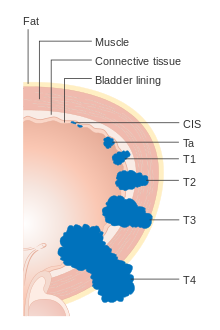
A bladder cancer is assigned a stage based on the TNM system, which is defined by the American Joint Committee on Cancer and used worldwide. A tumor is assigned three scores based on the extent of the primary tumor (T), its spread to nearby lymph nodes (N), and metastasis to distant sites (M). The T score represents the extent of the original tumor: Tis (for CIS tumors) or Ta (all others) for tumors that are confined to the innermost layer of the bladder; T1 for tumors that extend into the bladder's connective tissue; T2 for extension into the muscle; T3 for extension through the muscle into the surrounding fatty tissue; and T4 for extension fully outside the bladder. The N score represents spread to nearby lymph nodes: N0 for no spread; N1 for spread to a single nearby lymph node; N2 for spread to several nearby lymph nodes; N3 for spread to more distant lymph nodes outside the pelvis. The M score designates spread to more distant organs: M0 for a tumor that has not spread; M1 to one that has. The TNM scores are combined to determine the cancer case's stage on a scale of 0 to 4 and a higher stage means a more extensive cancer with a poorer prognosis.
Around 75% of cases are confined to the bladder at the time of diagnosis (T scores: Tis, Ta, or T1), and are called non-muscle-invasive bladder cancer (NMIBC). Around 18% have tumors that have spread into the bladder muscle (T2, T3, or T4), and are called muscle-invasive bladder cancer (MIBC). Around 3% have tumors that have spread to organs far from the bladder, and are called metastatic bladder cancer. Those with more extensive tumor spread tend to have a poorer prognosis.
| Stage | T | N | M | 5-year survival |
|---|---|---|---|---|
| 0 | Tis/Ta | N0 | M0 | 96% |
| 1 | T1 | N0 | M0 | 90% |
| 2 | T2 | N0 | M0 | 70% |
| 3 | T3 | N0 | M0 | 50% |
| 3 | Any T | N1-3 | M0 | 36% |
| 4 | Any T | Any N | M1 | 5% |
Treatment
Non-muscle-invasive bladder cancer
NMIBC is primarily treated by surgically removing all tumors by TURBT in the same procedure used to collect biopsy tissue for diagnosis. For those with a relatively low risk of tumors recurring, a single bladder injection of chemotherapy (mitomycin C, epirubicin, or gemcitabine) reduces the risk of tumors regrowing by about 40%. Those with higher risk are instead treated with bladder injections of the BCG vaccine (a live bacterial vaccine, traditionally used for tuberculosis), administered weekly for six weeks. This nearly halves the rate of tumor recurrence. Recurrence risk is further reduced by a series of "maintenance" BCG injections, given regularly for at least a year. Tumors that do not respond to BCG may be treated with the alternative immune stimulants nadofaragene firadenovec (sold as "Adstiladrin", a gene therapy that makes bladder cells produce an immunostimulant protein), nogapendekin alfa inbakicept ("Anktiva", a combination of immunostimulant proteins), or pembrolizumab ("Keytruda", an immune checkpoint inhibitor).

People whose tumors continue to grow are often treated with surgery to remove the bladder and surrounding organs, called radical cystectomy. The bladder, several adjacent lymph nodes, the lower ureters, and nearby internal genital organs – in men the prostate and seminal vesicles; in women the womb and part of the vaginal wall – are all removed. Surgeons construct a new way for urine to leave the body. The most common method is by ileal conduit, where a piece of the ileum (part of the small intestine) is removed and used to transport urine from the ureters to a new surgical opening (stoma) in the abdomen. Urine drains passively into an ostomy bag worn outside the body, which can be emptied regularly by the wearer. Alternatively, one can have a continent urinary diversion, where the ureters are attached to a piece of ileum that includes the valve between the small and large intestine; this valve naturally closes, allowing urine to be retained in the body rather than in an ostomy bag. The affected person empties the new urine reservoir serveral times each day by self-catheterization – passing a narrow tube through the stoma. Some can instead have the piece of ileum attached directly to the urethra, allowing the affected person to urinate through the urethra as they would pre-surgery – although without the original bladder nerves, they will no longer have the urge to urinate when the urine reservoir is full.
Radical cystectomy has both immediate and lifelong side effects. It is common for those recovering from surgery to experience gastrointestinal problems (29% of those who underwent radical cystectomy), infections (25%), and other issues with the surgical wound (15%). Around 25% of those who undergo the surgery end up readmitted to the hospital within 30 days; up to 2% die within 30 days of the surgery. Rerouting the ureters can also cause permanent metabolic issues. The piece of ileum used to reroute urine flow can absorb more ions from the urine than the original bladder would, resulting in metabolic acidosis (blood becoming too acidic), which can be treated with sodium bicarbonate. Shortening the small intestine can result in reduced vitamin B12 absorption, which can be treated with oral vitamin B12 supplementation. Issues with the new urine system can cause urinary retention, which can damage the ureters and kidneys and increase one's risk of urinary tract infection.
Those not well enough or unwilling to undergo radical cystectomy may instead benefit from further bladder injections of chemotherapy – mitomycin C, gemcitabine, docetaxel, or valrubicin – or intravenous injection of pembrolizumab. Around 1 in 5 people with NMIBC will eventually progress to MIBC.
Muscle-invasive bladder cancer
Most people with muscle-invasive bladder cancer are treated with radical cystectomy, which cures around half of those affected. Treating with chemotherapy prior to surgery (called "neoadjuvant therapy") using a cisplatin-containing drug combination (gemcitabine plus cisplatin; or methotrexate, vinblastine, doxorubicin, and cisplatin) improves survival an addition 5 to 10%.
Those with certain types of lower-risk disease may instead receive bladder-sparing therapy. People with just a single tumor at the back of the bladder can undergo partial cystectomy, with the tumor and surrounding area removed, and the bladder repaired. Those with no CIS or urinary blockage may undergo TURBT to remove visible tumors, followed by chemotherapy and radiotherapy; around two thirds of these people are cured. After treatment, surveillance tests – urine and blood tests, and MRI or CT scans – are done every three to six months to look for evidence that tumors may be recurring. Those who have retained their bladder also receive cystoscopies to look for additional bladder tumors. Recurrent bladder tumors are treated with radical cystectomy. Tumor recurrences elsewhere are treated as metastatic bladder cancer.
Metastatic disease
The standard of care for metastatic bladder cancer is combination treatment with the chemotherapy drugs cisplatin and gemcitabine. The average person on this combination survives around a year, though 15% experience remission, with survival over five years. Around half of those with metastatic bladder cancer are in too poor health to receive cisplatin. They instead receive the related drug carboplatin along with gemcitabine; the average person on this regimen survives around 9 months. Those whose disease responds to chemotherapy benefit from switching to immune checkpoint inhibitors pembrolizumab or atezolizumab ("Tecentriq") for long-term maintenance therapy. Immune checkpoint inhibitors are also commonly given to those whose tumors do not respond to chemotherapy, as well as those in too poor health to receive chemotherapy.
Those whose tumors continue to grow after platinum chemotherapy and immune checkpoint inhibitors can receive the antibody drug conjugate enfortumab vedotin ("Padcev", targets tumor cells with the protein nectin-4). Enfortumab vedotin in combination with pembrolizumab can also be used as a first-line therapy in place of chemotherapy. Those with genetic alterations that activate the proteins FGFR2 or FGFR3 (around 20% of those with metastatic bladder cancer) can also benefit from the FGFR inhibitor erdafitinib ("Balversa").
Bladder cancer that continues growing can be treated with second-line chemotherapies. Vinflunine is used in Europe, while paclitaxel, docetaxel, and pemetrexed are used in the United States; only a minority of those treated improve on these therapies.
Causes
Bladder cancer is caused by changes to the DNA of bladder cells that result in those cells growing uncontrollably. These changes can be random, or can be induced by exposure to toxic substances such as those from consuming tobacco. Genetic damage accumulates over many years, eventually disrupting the normal functioning of bladder cells and causing them to grow uncontrollably into a lump of cells called a tumor. Cancers cells accumulate further DNA changes as they multiply, which can allow the tumor to evade the immune system, resist regular cell death pathways, and eventually spread to distant body sites. The new tumors that form in various organs damage those organs, eventually causing the death of the affected person.
Smoking
Tobacco smoking is the main contributor to bladder cancer risk; around half of bladder cancer cases are estimated to be caused by smoking. Tobacco smoke contains carcinogenic molecules that enter the blood and are filtered by the kidneys into the urine. There they can cause damage to the DNA of bladder cells, eventually leading to cancer. Bladder cancer risk rises both with number of cigarettes smoked per day, and with duration of smoking habit. Those who smoke also tend to have an increased risk of treatment failure, metastasis, and death. The risk of developing bladder cancer decreases in those who quit smoking, falling by 30% after five years of smoking abstention. However, the risk will remain higher than those who have never smoked before. Because development of bladder cancer takes many years, it is not yet known if use of electronic cigarettes carries the same risk as smoking tobacco; however, those who use electronic cigarettes have higher levels of some urinary carcinogens than those who do not.
Occupational exposure
Up to 10% of bladder cancer cases are caused by workplace exposure to toxic chemicals. Exposure to certain aromatic amines, namely benzidine, beta-naphthylamine, and ortho-toluidine used in the metalworking and dye industries, can increase the risk of bladder cancer in metalworkers, dye producers, painters, printers, hairdressers, and textiles workers. The International Agency for Research on Cancer further classifies rubber processing, aluminum production, and firefighting as occupations that increase one's risk of developing bladder cancer. Exposure to arsenic – either through workplace exposure or through drinking water in places where arsenic naturally contaminates groundwater – is also commonly linked to bladder cancer risk.
Medical conditions
Chronic bladder infections can increase one's risk of developing bladder cancer. Most prominent is schistosomiasis, in which the eggs of the flatworm Schistosoma haematobium can become lodged in the bladder wall, causing chronic bladder inflammation and repeated bladder infections. In places with endemic schistosomiasis, up to 16% of bladder cancer cases are caused by prior Schistosoma infection. Worms can be cleared by treatment with praziquantel, which reduces bladder cancer cases in schistosomiasis endemic areas. Similarly, those with long-term indwelling catheters are at risk for repeated urinary tract infections, and have increased risk of developing bladder cancer.
Some medical treatments are also known to increase bladder cancer risk. As many as 16% of those treated with the chemotherapeutic cyclophosphamide go on to develop bladder cancer within 15 years of their treatment. Similarly, those treated with pelvic radiation (typically for prostate or cervical cancer) are at increased risk of developing bladder cancer five to 15 years after treatment. Long-term use of the medication pioglitazone for type 2 diabetes may increase bladder cancer risk.
Genetics
Bladder cancer does not typically run in families. Only 4% of those diagnosed with bladder cancer have a parent or sibling with the disease, potentially inheriting a gene syndrome associated with bladder cancer, for example:
- Mutations of the RB1 gene, while associated with 95% of Retinoblastoma cases, are also linked with bladder cancer.
- Mutation or genomic loss of the p16 protein, controlled by the CDKN2A tumor suppressor gene, is associated with 10-45% of bladder cancers, as well as glioma, esophageal cancer, head and neck cancer, non-small-cell lung cancer, breast cancer and colorectal cancer.
- Cowden disease is caused by mutations in the PTEN gene; see Cowden syndrome#Genetics. People with this syndrome have increased risk of developing several cancers, including bladder cancer.
- Lynch syndrome is caused by mutations in DNA mismatch repair genes MLH1, MSH2, MSH6 or PMS2; see main article Hereditary nonpolyposis colorectal cancer (HNPCC). People with this syndrome have increased risk of developing several cancers, including bladder and urinary tract cancers.
Large population studies have identified additional gene variants that each slightly increase bladder cancer risk. Most of these are variants in genes involved in metabolism of carcinogens (NAT2, GSTM1, and UGT1A6), controlling cell growth (TP63, CCNE1, MYC, and FGFR3), or repairing DNA damage (NBN, XRCC1 and 3, and ERCC2, 4, and 5).
Diet and lifestyle
Several studies have examined the impact of lifestyle factors on the risk of developing bladder cancer. A 2018 summary of evidence from the World Cancer Research Fund and American Institute for Cancer Research concluded that there is "limited, suggestive evidence" that consumption of tea, and a diet high in fruits and vegetables reduce a person's risk of developing bladder cancer. They also considered available data on exercise, body fat, and consumption of dairy, red meat, fish, grains, legumes, eggs, fats, soft drinks, alcohol, juices, caffeine, sweeteners, and various vitamins and minerals; for each they found insufficient data to link the lifestyle factor to bladder cancer risk. Several other studies have indicated a slight increased risk of developing bladder cancer in those who are overweight or obese, as well as a slight decrease in risk for those who undertake high levels of physical activity. Several studies have investigated a link between levels of fluid intake and bladder cancer risk – testing the theory that high fluid intake dilutes toxins in the urine and removes them through the body through more frequent urination – but have had inconsistent results, and a relationship remains unclear.
Pathophysiology
Despite arising from the same tissue, NMIBC and MIBC develop along distinct pathways and bear distinct genetic mutations. Most NMIBC tumors start as low-grade papillary (finger-like, projecting into the bladder) tumors. Mutations in cell growth pathways are common, most frequently activating mutations in FGFR3 (in up to 80% of NMIBC tumors). Mutations activating the growth pathway PI3K/AKT/mTOR pathway are also common, including activating mutations in PIK3CA (in around 30% of tumors) and ERBB2/ERBB3 (up to 15% of tumors), and loss of TSC1 (50% of tumors). Major regulators of chromatin (influences the expression of different genes) are inactivated in over 65% of NMIBC tumors.
MIBC often starts with low-lying, flat, high-grade tumors, that quickly spread beyond the bladder. These tumors have more genetic mutations and chromosomal abnormalities overall, with mutations more frequent than in any cancer but lung cancer and melanoma. Mutations that inactivate the tumor suppressor genes TP53 and RB are common, as are mutations in CDH1 (involved in metastasis) and VEGFR2 (involved in cell growth and metastasis).
Some genetic abnormalities are common to NMIBC and MIBC tumors. Around half of each have lost all or part of chromosome 9, which contains several regulators of tumor suppressor genes. Up to 80% of tumors have mutations in the gene TERT, which extends cells' telomeres to allow for extended replication.
Prognosis
Bladder cancer prognosis varies based on how widespread the cancer is at the time of diagnosis. For those with tumors confined to the innermost layer of the bladder (stage 0 disease), 96% are still alive five years from diagnosis. Those whose tumors have spread to nearby lymph nodes (stage 3 disease) have worse prognoses; 36% survive at least five years from diagnosis. Those with metastatic bladder cancer (stage 4 disease) have the most severe prognosis, with 5% alive five years from diagnosis.
Epidemiology

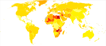
Around 500,000 people are diagnosed with bladder cancer each year, and 200,000 die of the disease. This makes bladder cancer the tenth most commonly diagnosed cancer, and the thirteenth cause of cancer deaths. Bladder cancer is most common in wealthier regions of the world, where exposure to certain carcinogens is highest. It is also common in places where schistosome infection is common, such as North Africa.
Bladder cancer is much more common in men than women; around 1.1% of men and 0.27% of women develop bladder cancer. This makes bladder cancer the sixth most common cancer in men, and the seventeenth in women. When women are diagnosed with bladder cancer, they tend to have more advanced disease and consequently a poorer prognosis. This difference in outcomes is attributed to numerous factors such as difference in carcinogen exposure, genetics, social factors and quality of care.
As with most cancers, bladder cancer is more common in older people; the average person with bladder cancer is diagnosed at age 73. 80% of those diagnosed with bladder cancer are 65 or older; 20% are 85 or older.
Veterinary medicine
Bladder cancer is relatively rare in domestic dogs and cats. In dogs, around 1% of diagnosed cancers are bladder cancer. Shetland sheepdogs, beagles, and various terriers are at increased risk relative to other breeds. Signs of a bladder tumor – blood in the urine, frequent urination, or trouble urinating – are common to other canine urinary conditions, and so diagnosis is often delayed. Urine tests can aid in diagnosis; they test for the protein bladder tumor antigen (high in bladder tumors) or mutations in the BRAF gene (present in around 80% of dogs with bladder or prostate cancer). Dogs with confirmed cancer are treated with COX-2 inhibitor drugs, such as piroxicam, deracoxib, and meloxicam. These drugs halt disease progression in around 50% of dogs, shrink tumors in around 12%, and eliminate tumors in around 6%. COX-2 inhibitors are often combined with chemotherapy drugs, most commonly mitoxantrone, vinblastine, or chlorambucil. Bladder cancer is much less common in cats than in dogs; treatment is similar to that of canine bladder cancer, with chemotherapy and COX-2 inhibitors commonly used.
References
- ^ Dyrskjøt et al. 2023, "Clinical Presentation".
- ^ Lenis, Lec & Chamie 2020, p. 1981.
- ^ Hahn 2022, "Clinical Presentation and Diagnostic Workup".
- ^ Hahn 2022, "Staging and Outcomes by Stage".
- "Symptoms of Metastatic Bladder Cancer". Cancer Research UK. 14 March 2023. Retrieved 11 November 2024.
- ^ "Tests for Bladder Cancer". American Cancer Society. 12 March 2024. Retrieved 8 August 2024.
- Dyrskjøt et al. 2023, "Diagnosis and Screening".
- ^ Ahmadi, Duddalwar & Daneshmand 2021, "Urine Cytology and Other Urine-Based Tumor Markers.
- ^ Ahmadi, Duddalwar & Daneshmand 2021, pp. 532–533.
- ^ Lenis, Lec & Chamie 2020, p. 1982.
- ^ Dyrskjøt et al. 2023, "Tissue-Based Diagnosis of Bladder Cancer".
- ^ "Types of Bladder Cancer". Cancer Research UK. 22 September 2024. Retrieved 26 August 2024.
- ^ "Stages of Bladder Cancer". Cancer Research UK. 22 September 2022. Retrieved 29 August 2024.
- ^ Hahn 2022, "Figure 86-2".
- ^ Hahn 2022, "Early-Stage Disease".
- Dyrskjøt et al. 2023, "TURBT and En Bloc Resection of Bladder Tumor".
- ^ Lenis, Lec & Chamie 2020, p. 1980.
- Dyrskjøt et al. 2023, "Intravesical Therapy for NMIBC".
- "Treatment of Bladder Cancer, Based on Stage and Other Factors". American Cancer Society. 1 May 2024.
- ^ Dyrskjøt et al. 2023, "Radical Cystectomy".
- "Surgery to Remove the Bladder (Cystectomy)". Cancer Research UK. 11 November 2022. Retrieved 16 September 2024.
- "Continent Urinary Diversion (Internal Pouch)". Cancer Research UK. 24 November 2022. Retrieved 17 September 2024.
- ^ Hahn 2022, "Muscle-Invasive Disease".
- "Bladder Reconstruction (Neobladder)". Cancer Reseaerch UK. 25 November 2024. Retrieved 17 September 2024.
- ^ Lenis, Lec & Chamie 2020, p. 1986.
- Teoh et al. 2022, p. 280.
- Dyrskjøt et al. 2023, "Perioperative Systemic Therapy".
- "Living as a Bladder Cancer Survivor". American Cancer Society. 12 March 2024. Retrieved 15 November 2024.
- ^ Compérat et al. 2022, pp. 1717–1718.
- ^ Hahn 2022, "Metastatic Disease".
- Dyrskjøt et al. 2023, "Systemic Therapy for Metastatic Bladder Cancer".
- ^ Lopez-Beltran et al. 2024, "Treatment of Metastatic Bladder Cancer".
- "Gilead Pulls Trodelvy's Approval in Bladder Cancer after Trial Flop, FDA Discussions". Fierce Pharma. 18 October 2024. Retrieved 15 November 2024.
- Smith et al. 2020, p. 1398.
- ^ "What Causes Bladder Cancer?". American Cancer Society. 12 March 2024. Retrieved 2 July 2024.
- "How Does Cancer Start". Cancer Research UK. 6 October 2023. Retrieved 24 October 2024.
- Hahn 2022, "Risk Factors".
- ^ Jubber et al. 2023, "3.2.1. Smoking".
- "Causes – Bladder Cancer". National Health Service. 1 July 2021. Retrieved 16 October 2024.
- Lobo et al. 2022, "3.2.1. Tobacco Smoking".
- Van Hoogstraten et al. 2023, pp. 291–292.
- Van Hoogstraten et al. 2023, pp. 292–293.
- Lobo et al. 2022, "3.2.2. Occupational carcinogen exposure".
- ^ Van Hoogstraten et al. 2023, p. 295.
- Dyrskjøt et al. 2023, "Occupational exposure".
- ^ Van Hoogstraten et al. 2023, p. 296.
- ^ Dyrskjøt et al. 2023, "Epidemiology".
- Lopez-Beltran et al. 2024, "Epidemiology".
- ^ Mossanen 2021, p. 449.
- ^ Hahn 2022, "Clinical Epidemiology and Risk Factors".
- Jubber et al. 2023, "3.2. Risk Factors for BC".
- ^ "Bladder Cancer Risk Factors". www.cancer.org. American Cancer Society. 12 March 2024. Retrieved 12 January 2025.
- ^ Mandigo AC, Tomlins SA, Kelly WK, Knudsen KE (19 January 2022). "Relevance of pRB Loss in Human Malignancies". Clinical Cancer Research. 28 (2): Table 1. doi:10.1158/1078-0432.CCR-21-1565. PMID 34407969. Retrieved 17 January 2025.
- ^ Van Hoogstraten et al. 2023, p. 293.
- "Diet, Nutrition, Physical Activity and Bladder Cancer" (PDF). World Cancer Research Fund, American Institute for Cancer Research. 2018. p. 20. Retrieved 17 October 2024.
- ^ Dyrskjøt et al. 2023, "Mechanisms/Pathophysiology".
- ^ Hahn 2022, "Molecular Biology".
- "WHO Disease and injury country estimates". World Health Organization. 2009. Archived from the original on 11 November 2009. Retrieved 11 November 2009.
- ^ Dyrskjøt et al. 2023, "Introduction".
- Marks P, Soave A, Shariat SF, Fajkovic H, Fisch M, Rink M (October 2016). "Female with bladder cancer: what and why is there a difference?". Translational Andrology and Urology. 5 (5): 668–682. doi:10.21037/tau.2016.03.22. PMC 5071204. PMID 27785424.
- Hahn 2022, "Introduction".
- Mossanen 2021, p. 448.
- ^ Burgess & DeRegis 2019, p. 314–317.
- Burgess & DeRegis 2019, p. 314–315.
- Griffin et al. 2020, "Introduction".
- Griffin et al. 2020, "3.5 Treatment".
Works cited
- Ahmadi H, Duddalwar V, Daneshmand S (June 2021). "Diagnosis and Staging of Bladder Cancer". Hematol Oncol Clin North Am. 35 (3): 531–541. doi:10.1016/j.hoc.2021.02.004. PMID 33958149.
- Burgess KE, DeRegis CJ (March 2019). "Urologic Oncology". Vet Clin North Am Small Anim Pract. 49 (2): 311–323. doi:10.1016/j.cvsm.2018.11.006. PMID 30635132.
- Compérat E, Amin MB, Cathomas R, Choudhury A, De Santis M, Kamat A, et al. (November 2022). "Current best practice for bladder cancer: a narrative review of diagnostics and treatments". Lancet. 400 (10364): 1712–1721. doi:10.1016/S0140-6736(22)01188-6. PMID 36174585.
- Dyrskjøt L, Hansel DE, Efstathiou JA, Knowles MA, Galsky MD, Teoh J, et al. (October 2023). "Bladder cancer". Nat Rev Dis Primers. 9 (1): 58. doi:10.1038/s41572-023-00468-9. PMC 11218610. PMID 37884563.
- Griffin MA, Culp WT, Giuffrida MA, Ellis P, Tuohy J, Perry JA, et al. (January 2020). "Lower urinary tract transitional cell carcinoma in cats: Clinical findings, treatments, and outcomes in 118 cases". J Vet Intern Med. 34 (1): 274–282. doi:10.1111/jvim.15656. PMC 6979092. PMID 31721288.
- Hahn NM (2022). "Chapter 86: Cancer of the Bladder and Urinary Tract". In Loscalzo J, Fauci A, Kasper D, et al. (eds.). Harrison's Principles of Internal Medicine (21st ed.). McGraw Hill. ISBN 978-1264268504.
- Jubber I, Ong S, Bukavina L, Black PC, Compérat E, Kamat AM, et al. (August 2023). "Epidemiology of Bladder Cancer in 2023: A Systematic Review of Risk Factors". Eur Urol. 84 (2): 176–190. doi:10.1016/j.eururo.2023.03.029. hdl:11573/1696425. PMID 37198015.
- Lenis AT, Lec PM, Chamie K (November 2020). "Bladder Cancer: A Review". JAMA. 324 (19): 1980–1991. doi:10.1001/jama.2020.17598. PMID 33201207.
- Lobo N, Afferi L, Moschini M, Mostafid H, Porten S, Psutka SP, et al. (December 2022). "Epidemiology, Screening, and Prevention of Bladder Cancer". Eur Urol Oncol. 5 (6): 628–639. doi:10.1016/j.euo.2022.10.003. PMID 36333236.
- Lopez-Beltran A, Cookson MS, Guercio BJ, Cheng L (February 2024). "Advances in diagnosis and treatment of bladder cancer". BMJ. 384: e076743. doi:10.1136/bmj-2023-076743. PMID 38346808.
- Mossanen M (June 2021). "The Epidemiology of Bladder Cancer". Hematol Oncol Clin North Am. 35 (3): 445–455. doi:10.1016/j.hoc.2021.02.001. PMID 33958144.
- Smith BA, Balar AV, Milowsky MI, Chen RC (2020). "Carcinoma of the Bladder". Abeloff's Clinical Oncology (6 ed.). Elsevier. pp. 1382–1400. ISBN 978-0-323-47674-4.
- Teoh JY, Kamat AM, Black PC, Grivas P, Shariat SF, Babjuk M (May 2022). "Recurrence mechanisms of non-muscle-invasive bladder cancer - a clinical perspective". Nat Rev Urol. 19 (5): 280–294. doi:10.1038/s41585-022-00578-1. PMID 35361927.
- Van Hoogstraten LM, Vrieling A, Van der Heijden AG, Kogevinas M, Richters A, Kiemeney LA (May 2023). "Global trends in the epidemiology of bladder cancer: challenges for public health and clinical practice". Nat Rev Clin Oncol. 20 (5): 287–304. doi:10.1038/s41571-023-00744-3. PMID 36914746.
External links
| Classification | D |
|---|---|
| External resources |
| Tumors of the urinary and genital systems | |||||||
|---|---|---|---|---|---|---|---|
| Kidney |
| ||||||
| Ureter | |||||||
| Bladder | |||||||
| Urethra | |||||||
| Other | |||||||