| This article needs additional citations for verification. Please help improve this article by adding citations to reliable sources. Unsourced material may be challenged and removed. Find sources: "Epimorphosis" – news · newspapers · books · scholar · JSTOR (December 2013) (Learn how and when to remove this message) |
Epimorphosis is defined as the regeneration of a specific part of an organism in a way that involves extensive cell proliferation of somatic stem cells, dedifferentiation, and reformation, as well as blastema formation. Epimorphosis can be considered a simple model for development, though it only occurs in tissues surrounding the site of injury rather than occurring system-wide. Epimorphosis restores the anatomy of the organism and the original polarity that existed before the destruction of the tissue and/or a structure of the organism. Epimorphosis regeneration can be observed in both vertebrates and invertebrates such as the common examples: salamanders, annelids, and planarians.
History
Thomas Hunt Morgan, an evolutionary biologist who also worked with embryology, argued that limb and tissue reformation bore many similarities to embryonic development. Building off of the work of German embryologist Wilhelm Roux, who suggested regeneration was two cooperative but distinct pathways instead of one, Morgan named the two parts of the regenerative process epimorphosis and morphallaxis. Specifically, Morgan wanted epimorphosis to specify the process of entirely new tissues being regrown from an amputation or similar injury, with morphallaxis being coined to describe regeneration that did not use cell proliferation, such as in hydra. The key difference between the two forms of regeneration is that epimorphosis involves cellular proliferation and blastema formation, whereas morphallaxis does not.
In vertebrates

In vertebrates, epimorphosis relies on blastema formation to proliferate cells into the new tissue. Through studies involving zebrafish fins, the toetips of mice, and limb regeneration in axolotls, researchers at the Polish Academy of Sciences found evidence for epimorphosis occurring in a variety of vertebrates, including instances of mammal epimorphosis.
Limb regeneration
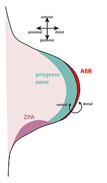
Limb regeneration occurs when a part of an organism is destroyed, and the organism must reform that structure. The general steps for limb regeneration are as follows: epidermis covers the wound which is called the wound healing process, the mesenchyme dedifferentiates into a blastema and a apical ectodermal cap forms, and the limb re-differentiates to form the full limb.
Processes in salamanders
Epidermal cells at the wound margins migrate to cover the wound and will become the wound epidermis. No scar tissue forms, as it would in mammals. The mesenchymal tissues of the limb stump secrete matrix metalloproteinases (MMPs). As the MMPs are secreted, the wound epithelium thickensand eventually becomes an apical ectodermal cap (AEC) that forms on the tip of the stump. This is similar to the embryonic apical ectodermal ridge, which forms during normal limb development. Under the AEC, the nerves near the site of the limb destroyed are degraded. The AEC causes the progress zone to re-establish; this means the cells under the AEC (including bone, cartilage, fibroblast cells, etc) dedifferentiate and become separated mesenchymal cells that form the blastema. Some tissues express specialized genes (like muscle cells) and so if there is damage to these tissues, the genes become downregulated and the proliferation genes are unregulated. The AEC also releases fibroblast growth factors (FGFs) (including FGF-4 and -8) that drive the development of the new limb, essentially resetting the limb back to its embryonic development stage. However, even though some of the limb cells are able to dedifferentiate, they are not able to fully dedifferentiate to the level of multipotent progenitor cells. During regeneration, only cartilage cells can form new cartilage tissue, only muscle cells can form new muscle tissue, and so on. The dedifferentiated cells still retain their original specification. To begin the physical formation of a new limb, regeneration occurs in a distal to proximal sequence. The distal part of the limb is established first, and then the distal part of the limb interacts with the original proximal part of the limb to form the intermediate portion of the limb known as intercalation.
In invertebrates
Periplaneta americana
The American cockroach is capable of regenerating limbs that have been damaged or destroyed, such as legs and antennae, as well parts of its compound eye. It does this with lectin—a protein made for binding proteins—named regenectin, which shares a family with other lipopolysaccharide (LPS) binding proteins. Regenectin carries both a regenerative and a system defense function, and it is produced by the cockroach's paracrine system to work with muscle reformation.
Capitella teleta
C. teleta is a segmented worm found in North America that is capable of regenerating posterior segments after amputation. This regeneration uses the interaction of several sets of Hox genes, as well as blastema formation. All of the Hox genes concerned in epimorphosis are present in the abdominal area of the worm, but not in the anterior portion. However, the genes do not, themselves, direct the anterior-posterior patterning of the worm's thorax.
Planaria vitta
P. vitta is a flatworm of genus Planaria that, when needed, can draw upon both morphallaxis and epimorphosis to regrow itself; in P. vitta, epimorphosis precedes morphallaxis and lasts about ten days. Planaria begin epimorphosis by the epidermis contracting immediately after the worm is cut at the head as a predator reactionary mechanism in order to decrease the surface area at the site of the cut. This mechanism activates the neoblasts which are totipotent stems cells which allows rhabdites to secrete materials to make a protective mucosal covering and epithelium to gather at the site through spreading of the cells rather than proliferation that occurs in vertebrates The dorsal and ventral epithelial cells then come to the site and become differentiated to begin regeneration. The polarity of the planaria can be reestablished through an anterior-posterior gradient through Wnt/β-catenin signaling pathway. Polarity can be described in planarians that the anterior part of the wound site will create a head of a planaria, and the posterior side will create the tail.
References
- "Medical Definition of EPIMORPHOSIS". www.merriam-webster.com. Retrieved 2018-02-19.
- Ribeiro RP, Bleidorn C, Aguado MT (March 2018). "Regeneration mechanisms in Syllidae (Annelida)". Regeneration. 5 (1): 26–42. doi:10.1002/reg2.98. PMC 5911452. PMID 29721325.
- Yokoyama H (January 2008). "Initiation of limb regeneration: the critical steps for regenerative capacity". Development, Growth & Differentiation. 50 (1): 13–22. doi:10.1111/j.1440-169X.2007.00973.x. PMID 17986260. S2CID 25299267.
- ^ Kubo T, Arai T (September 1996). "Insect Lectins and Epimorphosis". Trends in Glycoscience and Glycotechnology. 8 (43): 357–364. doi:10.4052/tigg.8.357.
- Sánchez Alvarado A, Tsonis PA (November 2006). "Bridging the regeneration gap: genetic insights from diverse animal models". Nature Reviews. Genetics. 7 (11): 873–84. doi:10.1038/nrg1923. PMID 17047686. S2CID 2978615.
- Sunderland ME (2010-05-01). "Regeneration: Thomas Hunt Morgan's window into development". Journal of the History of Biology. 43 (2): 325–61. doi:10.1007/s10739-009-9203-2. PMID 20665231. S2CID 24804711.
- ^ "Thomas Hunt Morgan's Definition of Regeneration: Morphallaxis and Epimorphosis". The Embryo Project Encyclopedia. Retrieved 2018-02-19.
- Summerbell D, Lewis JH, Wolpert L (August 1973). "Positional information in chick limb morphogenesis". Nature. 244 (5417): 492–6. Bibcode:1973Natur.244..492S. doi:10.1038/244492a0. PMID 4621272. S2CID 4166243.
- Conn PM (2017-06-20). Animal models for the study of human disease (Second ed.). London, United Kingdom. ISBN 978-0-12-809699-4. OCLC 992170104.
{{cite book}}: CS1 maint: location missing publisher (link) - Reddien PW, Sánchez Alvarado A (2004-10-08). "Fundamentals of planarian regeneration". Annual Review of Cell and Developmental Biology. 20 (1): 725–57. doi:10.1146/annurev.cellbio.20.010403.095114. PMID 15473858.
- Yokoyama H (January 2008). "Initiation of limb regeneration: the critical steps for regenerative capacity". Development, Growth & Differentiation. 50 (1): 13–22. doi:10.1111/j.1440-169X.2007.00973.x. PMID 17986260. S2CID 25299267.
- ^ Gilbert SF (2014). Developmental Biology (Tenth ed.). Sunderland, MA, USA: Sinauer Associates, Inc. pp. 571–573.
- ^ Yokoyama H (January 2008). "Initiation of limb regeneration: the critical steps for regenerative capacity". Development, Growth & Differentiation. 50 (1): 13–22. doi:10.1111/j.1440-169X.2007.00973.x. PMID 17986260. S2CID 25299267.
- Issues in Biological, Biochemical, and Evolutionary Sciences Research. Atlanta, GA: ScholarlyEditions. 2012. p. 464.
- Chernoff EA, Stocum DL (April 1995). "Developmental aspects of spinal cord and limb regeneration". Development, Growth and Differentiation. 37 (2): 133–147. doi:10.1046/j.1440-169x.1995.t01-1-00002.x. ISSN 0012-1592. PMID 37281907. S2CID 83821328.
- Nye HL, Cameron JA, Chernoff EA, Stocum DL (February 2003). "Regeneration of the urodele limb: a review". Developmental Dynamics. 226 (2): 280–94. doi:10.1002/dvdy.10236. PMID 12557206. S2CID 28442979.
- ^ Agata K, Saito Y, Nakajima E (February 2007). "Unifying principles of regeneration I: Epimorphosis versus morphallaxis". Development, Growth & Differentiation. 49 (2): 73–8. doi:10.1111/j.1440-169X.2007.00919.x. PMID 17335428.
- Kubo T, Arai T (September 1996). "Insect Lectins and Epimorphosis". Trends in Glycoscience and Glycotechnology. 8 (43): 357–364. doi:10.4052/tigg.8.357.
- Fröbius AC, Matus DQ, Seaver EC (2008-12-23). "Genomic organization and expression demonstrate spatial and temporal Hox gene colinearity in the lophotrochozoan Capitella sp. I". PLOS ONE. 3 (12): e4004. Bibcode:2008PLoSO...3.4004F. doi:10.1371/journal.pone.0004004. PMC 2603591. PMID 19104667.
- de Jong DM, Seaver EC (2016-02-19). "A Stable Thoracic Hox Code and Epimorphosis Characterize Posterior Regeneration in Capitella teleta". PLOS ONE. 11 (2): e0149724. Bibcode:2016PLoSO..1149724D. doi:10.1371/journal.pone.0149724. PMC 4764619. PMID 26894631.
- Newmark PA, Sánchez Alvarado A (March 2002). "Not your father's planarian: a classic model enters the era of functional genomics". Nature Reviews. Genetics. 3 (3): 210–9. doi:10.1038/nrg759. PMID 11972158. S2CID 28379017.
- ^ Chandebois R (August 1980). "The Dynamics of Wound Closure and Its Role in the Programming of Planarian Regeneration. II - Distalization". Development, Growth and Differentiation. 22 (4): 693–704. doi:10.1111/j.1440-169x.1980.00693.x. ISSN 0012-1592. PMID 37281333.
- Reddien PW, Sánchez Alvarado A (November 2004). "Fundamentals of planarian regeneration". Annual Review of Cell and Developmental Biology. 20 (1): 725–57. doi:10.1146/annurev.cellbio.20.010403.095114. PMID 15473858.
- Sánchez Alvarado A, Newmark PA (July 1998). "The use of planarians to dissect the molecular basis of metazoan regeneration". Wound Repair and Regeneration. 6 (4): 413–20. doi:10.1046/j.1524-475x.1998.60418.x. PMID 9824561. S2CID 8085897.
- ^ Morgan T (1901). "Regeneration". The American Historical Review. VII. doi:10.1086/ahr/17.4.809.