| External vertebral venous plexuses | |
|---|---|
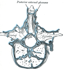
 Transverse section of a thoracic vertebra, showing the vertebral venous plexuses. Transverse section of a thoracic vertebra, showing the vertebral venous plexuses. | |
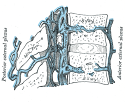
 Median sagittal section of two thoracic vertebrae, showing the vertebral venous plexuses. Median sagittal section of two thoracic vertebrae, showing the vertebral venous plexuses. | |
| Details | |
| Identifiers | |
| Latin | plexus venosi vertebrales externi |
| TA98 | A12.3.07.019 A12.3.07.020 |
| TA2 | 4951, 4952 |
| FMA | 12851 |
| Anatomical terminology[edit on Wikidata] | |
The external vertebral venous plexuses (extraspinal veins) consist of anterior and posterior plexuses which anastomose freely with each other. They are most prominent in the cervical region where they form anastomoses with the vertebral, occipital, and deep cervical veins.
- The anterior external vertebral venous plexuses are situated anteriorly to the vertebral bodies. They communicate with the basivertebral and intervertebral veins, and receive tributaries from the vertebral bodies.
- The posterior external vertebral venous plexuses are situated posterior to the vertebral laminae, around and the spinous, transverse, and articular processes. They form anastomoses with the internal vertebral venous plexuses, and drain to vertebral veins, posterior intercostal veins, and lumbar veins.
References
- ^ Standring, Susan (2020). Gray's Anatomy: The Anatomical Basis of Clinical Practice (42nd ed.). New York. p. 882. ISBN 978-0-7020-7707-4. OCLC 1201341621.
{{cite book}}: CS1 maint: location missing publisher (link) - Gray, Henry (1918). Gray's Anatomy (20th ed.). p. 668.
External links
- Atlas image: abdo_wall77 at the University of Michigan Health System - "Venous Drainage of the Vertebral Column"
| Veins of the thorax and vertebral column | |||||||||||||||
|---|---|---|---|---|---|---|---|---|---|---|---|---|---|---|---|
| Thorax |
| ||||||||||||||
| Vertebral column |
| ||||||||||||||
This cardiovascular system article is a stub. You can help Misplaced Pages by expanding it. |