| Vasoconstriction | |
|---|---|
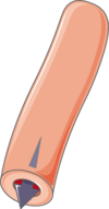
  Schematic depiction of relaxed vessel wall (left) and vasoconstriction (right) Schematic depiction of relaxed vessel wall (left) and vasoconstriction (right) | |
 Transmission electron micrograph showing vasoconstriction of a microvessel by pericytes and endothelial cells resulting in the deformation of an erythrocyte (E) Transmission electron micrograph showing vasoconstriction of a microvessel by pericytes and endothelial cells resulting in the deformation of an erythrocyte (E) | |
| Identifiers | |
| MeSH | D014661 |
| Anatomical terminology[edit on Wikidata] | |
Vasoconstriction is the narrowing of the blood vessels resulting from contraction of the muscular wall of the vessels, in particular the large arteries and small arterioles. The process is the opposite of vasodilation, the widening of blood vessels. The process is particularly important in controlling hemorrhage and reducing acute blood loss. When blood vessels constrict, the flow of blood is restricted or decreased, thus retaining body heat or increasing vascular resistance. This makes the skin turn paler because less blood reaches the surface, reducing the radiation of heat. On a larger level, vasoconstriction is one mechanism by which the body regulates and maintains mean arterial pressure.
Medications causing vasoconstriction, also known as vasoconstrictors, are one type of medicine used to raise blood pressure. Generalized vasoconstriction usually results in an increase in systemic blood pressure, but it may also occur in specific tissues, causing a localized reduction in blood flow. The extent of vasoconstriction may be slight or severe depending on the substance or circumstance. Many vasoconstrictors also cause pupil dilation. Medications that cause vasoconstriction include: antihistamines, decongestants, and stimulants. Severe vasoconstriction may result in symptoms of intermittent claudication.
General mechanism
The mechanism that leads to vasoconstriction results from the increased concentration of calcium (Ca ions) within vascular smooth muscle cells. However, the specific mechanisms for generating an increased intracellular concentration of calcium depends on the vasoconstrictor. Smooth muscle cells are capable of generating action potentials, but this mechanism is rarely utilized for contraction in the vasculature. Hormonal or pharmacokinetic components are more physiologically relevant. Two common stimuli for eliciting smooth muscle contraction are circulating epinephrine and activation of the sympathetic nervous system (through release of norepinephrine) that directly innervates the muscle. These compounds interact with cell surface adrenergic receptors. Such stimuli result in a signal transduction cascade that leads to increased intracellular calcium from the sarcoplasmic reticulum through IP3-mediated calcium release, as well as enhanced calcium entry across the sarcolemma through calcium channels. The rise in intracellular calcium complexes with calmodulin, which in turn activates myosin light-chain kinase. This enzyme is responsible for phosphorylating the light chain of myosin to stimulate cross-bridge cycling.
Once elevated, the intracellular calcium concentration is returned to its normal concentration through a variety of protein pumps and calcium exchangers located on the plasma membrane and sarcoplasmic reticulum. This reduction in calcium removes the stimulus necessary for contraction, allowing for a return to baseline.
Causes
Factors that trigger vasoconstriction can be exogenous or endogenous in origin. Ambient temperature is an example of exogenous vasoconstriction. Cutaneous vasoconstriction will occur because of the body's exposure to the severe cold. Examples of endogenous factors include the autonomic nervous system, circulating hormones, and intrinsic mechanisms inherent to the vasculature itself (also referred to as the myogenic response).
Exposure to water causes vasoconstriction near the skin, which in turn causes water-immersion wrinkling.
Examples
Examples include stimulants, amphetamines, and antihistamines. Many are used in medicine to treat hypotension and as topical decongestants. Vasoconstrictors are also used clinically to increase blood pressure or to reduce local blood flow. Vasoconstrictors mixed with local anesthetics are used to increase the duration of local anesthesia by constricting the blood vessels, thereby safely concentrating the anesthetic agent for an extended duration, as well as reducing hemorrhage.
The routes of administration vary. They may be both systemic and topical. For example, pseudoephedrine is taken orally and phenylephrine is topically applied to the nasal passages or eyes. Examples include:
Endogenous
Vasoconstriction is a procedure of the body that averts orthostatic hypotension. It is part of a body negative feedback loop in which the body tries to restore homeostasis (maintain constant internal environment).
For example, vasoconstriction is a hypothermic preventative in which the blood vessels constrict and blood must move at a higher pressure to actively prevent a hypoxic reaction. ATP is used as a form of energy to increase this pressure to heat the body. Once homeostasis is restored, the blood pressure and ATP production regulates. Vasoconstriction also occurs in superficial blood vessels of warm-blooded animals when their ambient environment is cold; this process diverts the flow of heated blood to the center of the animal, preventing the loss of heat.
| Vasoconstrictor | Receptor (↑ = opens. ↓ = closes) On vascular smooth muscle cells if not otherwise specified |
Transduction (↑ = increases. ↓ = decreases) |
|---|---|---|
| Stretch | ↑Stretch-activated ion channels | depolarization -->
|
| ATP (intracellular) | ↓ATP-sensitive K channel | |
| ATP (extracellular) | ↑P2X receptor | ↑Ca |
| NPY | NPY receptor | Activation of Gi --> ↓cAMP --> ↓PKA activity --> ↓phosphorylation of MLCK --> ↑MLCK activity --> ↑phosphorylation of MLC (calcium-independent) |
| adrenergic agonists e.g., epinephrine, norepinephrine, and dopamine |
↑α1 adrenergic receptor | Activation of Gq --> ↑PLC activity --> ↑IP3 and DAG --> activation of IP3 receptor in SR --> ↑intracellular Ca |
| thromboxane | ↑thromboxane receptor | |
| endothelin | ↑endothelin receptor ETA | |
| angiotensin II | ↑Angiotensin receptor 1 |
|
| open VDCCs --> ↑intracellular Ca | ||
| Asymmetric dimethylarginine | Reduced production of nitric oxide | |
| Antidiuretic hormone (ADH or Vasopressin) | Arginine vasopressin receptor 1 (V1) on smooth muscle cells | Activation of Gq --> ↑PLC activity --> ↑IP3 and DAG --> activation of IP3 receptor in SR --> ↑intracellular Ca |
| Arginine vasopressin receptor on endothelium | Endothelin production | |
|
Various receptors on endothelium | Endothelin production |
Pathology
Vasoconstriction can be a contributing factor to erectile dysfunction. An increase in blood flow to the penis causes an erection.
Improper vasoconstriction may also play a role in secondary hypertension.
To summarize, vasoconstriction is a physiological process that involves the narrowing of blood vessels, particularly arteries and arterioles, resulting in a reduction of blood flow to specific tissues or organs. This phenomenon is primarily regulated by the contraction of smooth muscle cells within the vessel walls. Several factors contribute to vasoconstriction, including the release of vasoconstrictor substances such as endothelin and angiotensin II, both of which play crucial roles in the modulation of vascular tone.
Additionally, sympathetic nervous system activation, triggered by stress or other stimuli, prompts the release of norepinephrine, a neurotransmitter that induces vasoconstriction by binding to alpha-adrenergic receptors on smooth muscle cells. The narrowing of blood vessels leads to an increase in peripheral resistance, thereby elevating blood pressure. While vasoconstriction is a normal and essential regulatory mechanism for maintaining blood pressure and redistributing blood flow during various physiological processes, its dysregulation can contribute to pathological conditions. Chronic vasoconstriction is associated with hypertension, a major risk factor for cardiovascular diseases such as heart attack and stroke. Moreover, impaired blood flow resulting from abnormal vasoconstriction may contribute to tissue ischemia, which can be observed in conditions like Raynaud's disease. Understanding the pathology of vasoconstriction is crucial for developing targeted therapeutic strategies to manage conditions associated with abnormal vascular tone.
See also
- Addison's disease
- Inotrope
- Hypertension
- Nitric oxide
- Pheochromocytoma
- Shock
- Vasodilation
- Postural orthostatic tachycardia syndrome
- Hemostasis
References
- "Medihaler Ergotamine". drugs.com. Retrieved 2016-05-20.
-
Michael P. Walsh; et al. (2005-08-01) . "Thromboxane A2-induced contraction of rat caudal arterial smooth muscle involves activation of Ca entry and Casensitization: Rho-associated kinase-mediated phosphorylation of MYPT1 at Thr-855, but not Thr-697". Biochem J. 389 (3): 763–774. doi:10.1042/BJ20050237. PMC 1180727. PMID 15823093.
These results suggest that U-46619 elicits contraction of rat caudal arterial smooth muscle by activating Ca entry from the extracellular space, which may or may not involve Ca-induced Ca release from the SR (sarcoplasmic reticulum). ... A key step in the contractile response to U-46619 appears to be the entry of extracellular Ca, since it was abolished by removal of extracellular Ca (Figure 2A). ... In the rat caudal artery, U-46619-mediated contractile responses have an absolute requirement for Ca, which enters from the extracellular pool, is independent of intracellular Ca stores and is blocked by ROK inhibition.
- Butler; Siegman (December 1998). "Control of cross‐bridge cycling by myosin light chain phosphorylation in mammalian smooth muscle". Acta Physiologica Scandinavica. 164 (4): 389–400. doi:10.1046/j.1365-201x.1998.00450.x. PMID 9887963.
- Yagiela JA (1995). "Vasoconstrictor agents for local anesthesia". Anesth Prog. 42 (3–4): 116–20. PMC 2148913. PMID 8934977.
- Moodley, D. S. (May 2017). "Local anaesthetics in dentistry - Part 3: Vasoconstrictors in local anaesthetics". South African Dental Journal. 72 (4): 176–178. hdl:10566/3893.
- Salerno, Stephen M.; Jackson, Jeffrey L.; Berbano, Elizabeth P. (8 August 2005). "Effect of Oral Pseudoephedrine on Blood Pressure and Heart Rate: A Meta-analysis". Archives of Internal Medicine. 165 (15): 1686–1694. doi:10.1001/archinte.165.15.1686. PMID 16087815.
- Horak, Friedrich; Zieglmayer, Petra; Zieglmayer, René; Lemell, Patrick; Yao, Ruji; Staudinger, Heribert; Danzig, Melvyn (February 2009). "A placebo-controlled study of the nasal decongestant effect of phenylephrine and pseudoephedrine in the Vienna Challenge Chamber". Annals of Allergy, Asthma & Immunology. 102 (2): 116–120. doi:10.1016/s1081-1206(10)60240-2. PMID 19230461.
- Halberstadt, Adam L. (2017). "Pharmacology and Toxicology of N-Benzylphenethylamine ('NBOMe') Hallucinogens". Neuropharmacology of New Psychoactive Substances (NPS). Current Topics in Behavioral Neurosciences. Vol. 32. pp. 283–311. doi:10.1007/7854_2016_64. ISBN 978-3-319-52442-9. PMID 28097528.
- Echeverri, Darío; Montes, Félix R.; Cabrera, Mariana; Galán, Angélica; Prieto, Angélica (25 August 2010). "Caffeine's Vascular Mechanisms of Action". International Journal of Vascular Medicine. 2010: 1–10. doi:10.1155/2010/834060. PMC 3003984. PMID 21188209.
- Laccourreye, O.; Werner, A.; Giroud, J.-P.; Couloigner, V.; Bonfils, P.; Bondon-Guitton, E. (2015). "Benefits, limits and danger of ephedrine and pseudoephedrine as nasal decongestants". European Annals of Otorhinolaryngology, Head and Neck Diseases. 132 (1): 31–34. doi:10.1016/j.anorl.2014.11.001. PMID 25532441.
- Hwang, Kun; Son, Ji Soo; Ryu, Woo Kyung (November 2018). "Smoking and Flap Survival". Plastic Surgery (Oakville, Ont.). 26 (4): 280–285. doi:10.1177/2292550317749509. ISSN 2292-5503. PMC 6236508. PMID 30450347.
- ^ Unless else specified in box, then ref is: Walter F. Boron (2005). Medical Physiology: A Cellular And Molecular Approaoch. Elsevier/Saunders. ISBN 1-4160-2328-3. Page 479
- ^ Rod Flower; Humphrey P. Rang; Maureen M. Dale; Ritter, James M. (2007). Rang & Dale's pharmacology. Edinburgh: Churchill Livingstone. ISBN 978-0-443-06911-6.
- Walter F. Boron (2005). Medical Physiology: A Cellular And Molecular Approach. Elsevier/Saunders. ISBN 1-4160-2328-3. Page 771
- Richard Milsten and Julian Slowinski, The sexual male, bc, main point W.W. Norton Company, New York, London (1999) ISBN 0-393-04740-7
- Swenson, Erik R. (June 2013). "Hypoxic Pulmonary Vasoconstriction". High Altitude Medicine & Biology. 14 (2): 101–110. doi:10.1089/ham.2013.1010. PMID 23795729.
- Temprano, Katherine K. (March 2016). "A Review of Raynaud's Disease". Missouri Medicine. 113 (2): 123–126. PMC 6139949. PMID 27311222.
External links
- Definition of Vasoconstriction on HealthScout
- Disdier, Patrick; Granel, Brigitte; Serratrice, Jacques; Constans, Joël; Michon-Pasturel, Ulrique; Hachulla, Eric; Conri, Claude; Devulder, Bernard; Swiader, Laure; Piquet, Philippe; Branchereau, Alain; Jouglard, Jacqueline; Moulin, Guy; Weiller, Pierre-Jean (January 2001). "Cannabis Arteritis Revisited: Ten New Case Reports". Angiology. 52 (1): 1–5. doi:10.1177/000331970105200101. PMID 11205926. S2CID 26030253.
- Are coronary heart disease and peripheral arterial disease associated with tobacco or cannabis consumption
| Physiology of the cardiovascular system | |||||||||||||
|---|---|---|---|---|---|---|---|---|---|---|---|---|---|
| Heart |
| ||||||||||||
| Vascular system/ hemodynamics |
| ||||||||||||